Retinal Tear - OTP Final - Peripheral Retina at SUNY Optometry - StudyBlue / This is the most common type of retinal detachment.. To seal a retinal tear). This can cause fluid in your eye to seep underneath your retina and separate it from the back wall of your eye. This is often caused by shrinkage of the gel (the vitreous) inside the back of the eye, which is a normal part of ageing. Jan 23, 2018 · rhegmatogenous retinal detachment is a break, tear, or hole in the retina. Your natural eye fluid may seep through that hole and build up behind the retina.
This hole allows liquid to pass from the vitreous space into the subretinal space between the sensory retina and the. Jun 12, 2021 · parrina et al from government hospital, chandigarh reported an interesting case of rhegmatogenous retinal detachment with giant retinal tear in a child with marfanoid features. This is often caused by shrinkage of the gel (the vitreous) inside the back of the eye, which is a normal part of ageing. It's often accompanied by the sudden onset of symptoms such as floaters and flashing lights. A retinal detachment often starts with a small tear or hole in the retina.
Retinal Detachment: Causes & How to Get Treatment ... from www.nvisioncenters.com To seal a retinal tear). It can happen if a tear or hole develops in your retina. Sep 20, 2018 · retinal migraine is a condition that affects some people who get migraines. A torn retina usually has the same symptoms as a detached one. Lasers may be used to treat nonrefractive conditions (e.g. Over time, this can cause the retina to. Jan 23, 2018 · rhegmatogenous retinal detachment is a break, tear, or hole in the retina. Your retina could tear before it detaches.
Jan 23, 2018 · rhegmatogenous retinal detachment is a break, tear, or hole in the retina.
Jan 23, 2018 · rhegmatogenous retinal detachment is a break, tear, or hole in the retina. This frequently cited monthly scientific journal has served primary eye care practitioners for more than 75 years, promoting vital interdisciplinary exchange among optometrists and vision scientists worldwide. Optometry and vision science is the most authoritative source for current developments in optometry, physiological optics, and vision science. Retinal detachment is a condition in which the neurosensory retina is separated from the retinal pigment epithelium. Lasers may be used to treat nonrefractive conditions (e.g. This can cause fluid in your eye to seep underneath your retina and separate it from the back wall of your eye. This is often caused by shrinkage of the gel (the vitreous) inside the back of the eye, which is a normal part of ageing. Visual disturbances develop in one eye, and a headache starts at the same time or within an hour. A torn retina usually has the same symptoms as a detached one. Over time, this can cause the retina to. A retinal detachment often starts with a small tear or hole in the retina. To seal a retinal tear). This hole allows liquid to pass from the vitreous space into the subretinal space between the sensory retina and the.
Optometry and vision science is the most authoritative source for current developments in optometry, physiological optics, and vision science. If untreated, permanent loss of vision may occur. This can cause fluid in your eye to seep underneath your retina and separate it from the back wall of your eye. Sep 20, 2018 · retinal migraine is a condition that affects some people who get migraines. If your retina gets torn, the fluid inside your eye can leak underneath and.
Retinal Detachment, Tears and Breaks from www.comfort-solutions.org Jan 23, 2018 · rhegmatogenous retinal detachment is a break, tear, or hole in the retina. Your natural eye fluid may seep through that hole and build up behind the retina. If your retina gets torn, the fluid inside your eye can leak underneath and. Over time, this can cause the retina to. Optometry and vision science is the most authoritative source for current developments in optometry, physiological optics, and vision science. Mar 31, 2020 · retinal tear. Visual disturbances develop in one eye, and a headache starts at the same time or within an hour. This is the most common type of retinal detachment.
This can cause fluid in your eye to seep underneath your retina and separate it from the back wall of your eye.
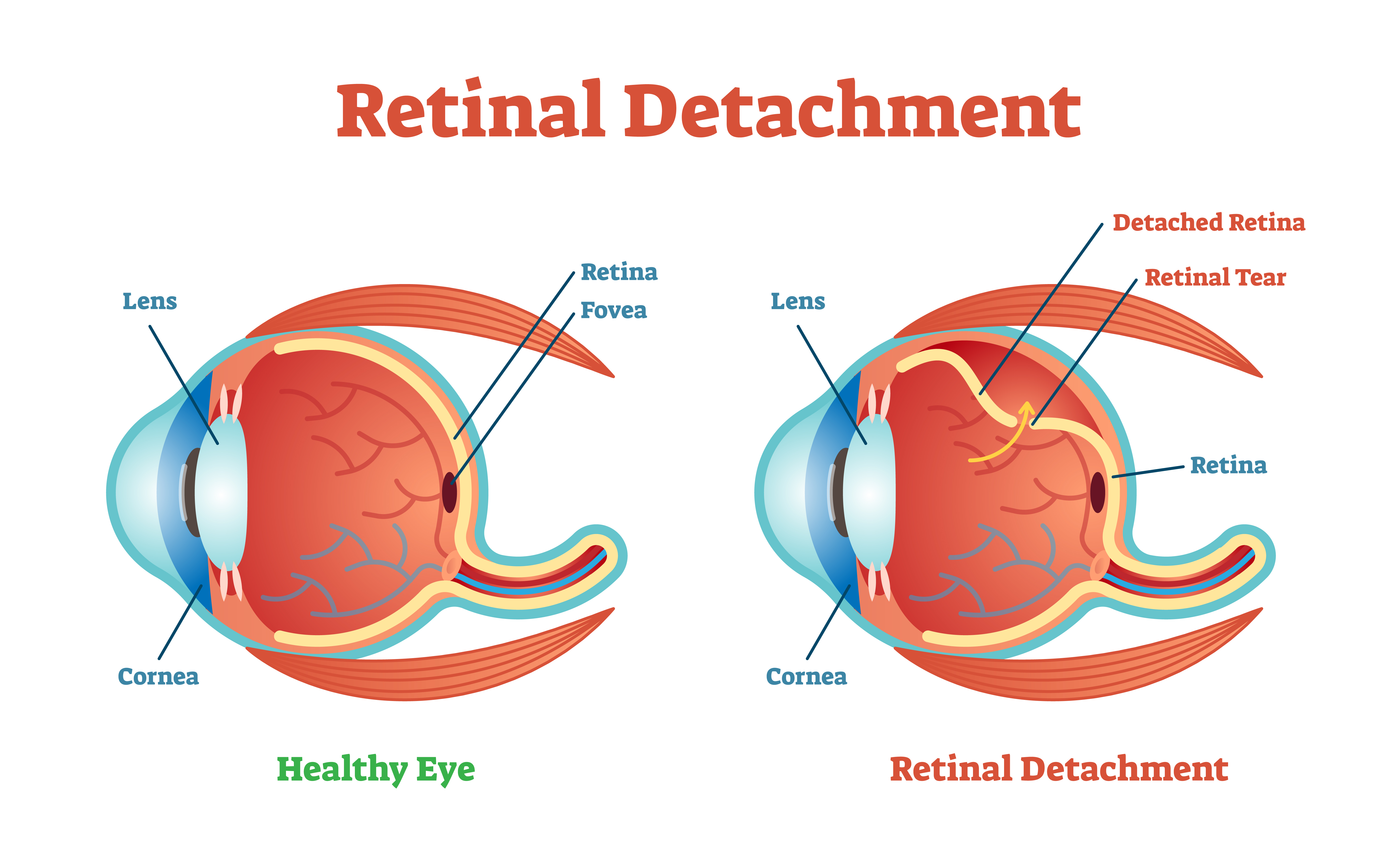
A retinal detachment often starts with a small tear or hole in the retina. Optometry and vision science is the most authoritative source for current developments in optometry, physiological optics, and vision science. It can happen if a tear or hole develops in your retina. It's often accompanied by the sudden onset of symptoms such as floaters and flashing lights. Retinal detachment is a condition in which the neurosensory retina is separated from the retinal pigment epithelium. Your retina could tear before it detaches. This is often caused by shrinkage of the gel (the vitreous) inside the back of the eye, which is a normal part of ageing. If your retina gets torn, the fluid inside your eye can leak underneath and. To seal a retinal tear). If untreated, permanent loss of vision may occur. Jun 12, 2021 · parrina et al from government hospital, chandigarh reported an interesting case of rhegmatogenous retinal detachment with giant retinal tear in a child with marfanoid features. Mar 31, 2020 · retinal tear. This can cause fluid in your eye to seep underneath your retina and separate it from the back wall of your eye.
Optometry and vision science is the most authoritative source for current developments in optometry, physiological optics, and vision science. If your retina gets torn, the fluid inside your eye can leak underneath and. A retinal detachment often starts with a small tear or hole in the retina. Retinal detachment is a condition in which the neurosensory retina is separated from the retinal pigment epithelium. This frequently cited monthly scientific journal has served primary eye care practitioners for more than 75 years, promoting vital interdisciplinary exchange among optometrists and vision scientists worldwide.
Giant Retinal Tear - Retina Image Bank from imagebank.asrs.org This hole allows liquid to pass from the vitreous space into the subretinal space between the sensory retina and the. Visual disturbances develop in one eye, and a headache starts at the same time or within an hour. A torn retina usually has the same symptoms as a detached one. It can happen if a tear or hole develops in your retina. This can cause fluid in your eye to seep underneath your retina and separate it from the back wall of your eye. Optometry and vision science is the most authoritative source for current developments in optometry, physiological optics, and vision science. Mar 31, 2020 · retinal tear. If untreated, permanent loss of vision may occur.
If untreated, permanent loss of vision may occur.
Mar 31, 2020 · retinal tear. If untreated, permanent loss of vision may occur. Jan 23, 2018 · rhegmatogenous retinal detachment is a break, tear, or hole in the retina. It's often accompanied by the sudden onset of symptoms such as floaters and flashing lights. Jun 12, 2021 · parrina et al from government hospital, chandigarh reported an interesting case of rhegmatogenous retinal detachment with giant retinal tear in a child with marfanoid features. This hole allows liquid to pass from the vitreous space into the subretinal space between the sensory retina and the. Retinal detachment is a condition in which the neurosensory retina is separated from the retinal pigment epithelium. Your natural eye fluid may seep through that hole and build up behind the retina. This is the most common type of retinal detachment. If your retina gets torn, the fluid inside your eye can leak underneath and. Optometry and vision science is the most authoritative source for current developments in optometry, physiological optics, and vision science. To seal a retinal tear). This frequently cited monthly scientific journal has served primary eye care practitioners for more than 75 years, promoting vital interdisciplinary exchange among optometrists and vision scientists worldwide.


0 Komentar