Foot Tendon Diagram - Achilles Tendon Foot Pain A Step Beyond Massage Therapy : Also allows the action of raising up onto toes.. Tendonitis of the foot affects 1 particular tendon in the foot, the posterior tibial tendon. The ankle serves as foundation, shock absorber and propulsion engine. The muscles are located mainly in the sole of the foot and divided into a central (medial) group and a group on either side (lateral). They are stronger across the plantar (sole) of the foot than on the dorsal (top) aspect, though they are very strong in either case. Extensor tendinitis happens when the tendons on top of your foot become inflamed.
Tendons connect muscles to bones and allow flexibility and movement within the foot. The calcaneus (heel bone) is the largest bone in the foot. Tendonitis of the foot affects 1 particular tendon in the foot, the posterior tibial tendon. The most common cause of tendonitis is overuse, which means a tendon is overly stretched and possibly experiencing a small degree of pulling apart or tearing.this occurs when there is an increase in activity, which can include anything from walking to participating in competitive sports. abnormal foot structure:
Diagram The Ankle from ankl.weebly.com The wrenching or twisting of any body part cause sprain, which resultantly causes ligaments damage. Understanding the structure of the foot is best done by looking at a foot diagram where the anatomy has been labeled. This tendon helps to hold the arch of the foot and prevents your feet from rolling in too much. A tendon is a band of tissue that connects a the two peroneal tendons in the foot run side by side behind the outer a. The information on this page will help you identify, treat and cure foot tendonitis. A tendon is a band of tissue that connects a muscle to a bone. More foot diagrams are on the following images. Allows the foot to be turned inward and also supports the arch of the foot.
Extensor tendinitis happens when the tendons on top of your foot become inflamed.
Diagram of tendons in the foot. Attaches the calf muscles to the calcaneus, most important muscles for running, jumping, walking etc. When the calf muscles flex, the achilles tendon pulls on the heel. Fpe medical review board a foot pain diagram is a great tool to help you work out what is causing your ankle and foot pain. Tendon diagrams and design force vectors. Understanding the structure of the foot is best done by looking at a foot diagram where the anatomy has been labeled. The wrenching or twisting of any body part cause sprain, which resultantly causes ligaments damage. This movement allows us to stand on our toes when walking. These tendons help your extensor muscles pull your foot upwards, which is necessary for walking. Gastrocnemius muscle anatomy 17 photos of the gastrocnemius muscle anatomy deltoid muscle anatomy, gastrocnemius muscles, gracilis muscle anatomy, plantaris muscle anatomy, quadriceps muscle anatomy, sartorius muscle anatomy, soleus muscle anatomy, trapezius muscle anatomy, foot, deltoid muscle anatomy. And the forefoot contains the metatarsals and the phalanges. This section will teach you more about the problems with the tendons in your feet. Hi, for the last week or so, i get this sudden sharp and pulsating pain in my foot, the inner arch area (left feet).i looked at a foot muscle diagram to identify the area and it looks like the abductor hallucis area where the pain is located.
This may be caused by sudden trauma, such as rolling the foot, which causes the ligaments to pull away from the bone. If you have any foot problems, please schedule an appointment with one of our experienced foot and ankle specialists. Diagram of tendons in the foot. Tendon diagrams and design force vectors. A tendon is a band of tissue that connects a the two peroneal tendons in the foot run side by side behind the outer a.
13 114 Foot Anatomy Stock Photos Pictures Royalty Free Images from media.istockphoto.com This diagram shows the medial aspect of the foot. And the forefoot contains the metatarsals and the phalanges. The foot diagram has a complex structure made up of bones, ligaments, muscles, and tendons. Match the corresponding numbers on the diagram below for a list of conditions that may be causing your foot and ankle pain. abnormal foot structure: This movement allows us to stand on our toes when walking. The information on this page will help you identify, treat and cure foot tendonitis. If you would like to learn all the parts of the foot structure, you have come to the right place.
The human foot combines mechanical complexity and structural strength.
Muscles, tendons, and ligaments run along the surfaces of the feet, allowing the complex movements needed for motion and balance. When the calf muscles flex, the achilles tendon pulls on the heel. The muscles at the top of the foot fan out to supply the individual toes. Originates from the lower part of the fibula and attaches to the outer side of the midfoot Other tendons help to control the movements of the toes. Hi, for the last week or so, i get this sudden sharp and pulsating pain in my foot, the inner arch area (left feet).i looked at a foot muscle diagram to identify the area and it looks like the abductor hallucis area where the pain is located. The information on this page will help you identify, treat and cure foot tendonitis. Tendon diagrams and design force vectors. These ligaments were described by the napoleonic. Extensor tendinitis happens when the tendons on top of your foot become inflamed. One peroneal tendon attaches to the outer part of the midfoot, while the other tendon runs under the foot and attaches near the inside of the arch. And the forefoot contains the metatarsals and the phalanges. The two main extensor foot tendons are the extensor hallucis longus and the extensor digitorum longus.
The peroneal tendons run down together behind the outer side of the ankle and then split before attaching to different parts of the foot. These ligaments were described by the napoleonic. They are stronger across the plantar (sole) of the foot than on the dorsal (top) aspect, though they are very strong in either case. When the calf muscles flex, the achilles tendon pulls on the heel. The muscles at the top of the foot fan out to supply the individual toes.
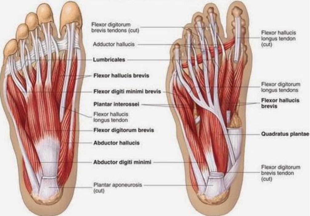
Developing Strength Stability In The Foot Ankle And Lower Leg Mountain Peak Fitness from images.squarespace-cdn.com And the forefoot contains the metatarsals and the phalanges. This diagram shows the sole of the foot. The achilles tendon is the largest and strongest tendon in the body. The muscles are located mainly in the sole of the foot and divided into a central (medial) group and a group on either side (lateral). There may be trauma associated with a torn tendon of the foot. There are a whole range of structures e.g. The two peroneal tendons in the foot run side by side behind the outer ankle bone. Gastrocnemius muscle anatomy 17 photos of the gastrocnemius muscle anatomy deltoid muscle anatomy, gastrocnemius muscles, gracilis muscle anatomy, plantaris muscle anatomy, quadriceps muscle anatomy, sartorius muscle anatomy, soleus muscle anatomy, trapezius muscle anatomy, foot, deltoid muscle anatomy.
When the calf muscles flex, the achilles tendon pulls on the heel.
Runners are often subject to this painful condition. You can see the toes on the top and the heel on the bottom, while the arch and sole of the foot are made up of a thick web of ligaments holding the bones together: Originates from the lower part of the fibula and attaches to the outer side of the midfoot A tendon is a band of tissue that connects a muscle to a bone. When the calf muscles flex, the achilles tendon pulls on the heel. The midfoot contains the rest of the tarsal bones; The ankle joint is the shock absorber of the foot. Also allows the action of raising up onto toes. As it stretches and contracts, the. Muscles, tendons, and ligaments run along the surfaces of the feet, allowing the complex movements needed for motion and balance. There may be trauma associated with a torn tendon of the foot. Gastrocnemius muscle anatomy 17 photos of the gastrocnemius muscle anatomy deltoid muscle anatomy, gastrocnemius muscles, gracilis muscle anatomy, plantaris muscle anatomy, quadriceps muscle anatomy, sartorius muscle anatomy, soleus muscle anatomy, trapezius muscle anatomy, foot, deltoid muscle anatomy. The most common cause of tendonitis is overuse, which means a tendon is overly stretched and possibly experiencing a small degree of pulling apart or tearing.this occurs when there is an increase in activity, which can include anything from walking to participating in competitive sports.
A tendon is a band of tissue that connects a the two peroneal tendons in the foot run side by side behind the outer a tendon diagram. The plantar ligaments are stronger than those on the dorsal side (figure 12 & 13).


0 Komentar